
To Confirm and Measure Fatty Liver
Ultrasound is recommended for the first look at suspected fatty liver, and MRI-PDFF when a precise fat measurement is needed. It can show whether fat is present and how much.
AI Summary MRI-PDFF is a special liver MRI that measures fat as a percentage, using magnets and radio waves with no radiation. Doctors order it to confirm fatty liver, which starts at about 5% fat, and to track changes over time. It does not measure scarring. Results are read with blood tests and other imaging. Lifestyle changes such as weight loss come first, and a biopsy remains the standard for diagnosing MASH.
Source: www.liverdirectory.com
Medically Reviewed.
MRI-PDFF stands for magnetic resonance imaging–proton density fat fraction. It is a special type of liver MRI that reports fat as a percentage of the liver. The scan uses magnets and radio waves, so it involves no radiation.
Doctors use it to confirm fatty liver disease, to see how much fat is present, and to check whether fat goes down over time. It measures fat only. It does not measure scarring, so your care team reads it with blood tests and other tests.

 Liver Health AI Assistant

Ask Aimy
Aimy answers from LiverDirectory’s reviewed health information — helping patients make sense of liver-health terms, prepare for specialist visits, and explore what to look up next.
LIVER FAT MRI BASICS
MRI-PDFF is a liver MRI method that measures fat. An MRI scanner uses powerful magnets and radio waves to create many thin images, called slices, of the liver. With PDFF, the scanner's software turns those images into a fat measurement, shown as a percentage.
The percentage tells you how much of the liver is fat. A small amount of fat is normal. About 5% or more is the point where fatty liver is present.
Fatty liver disease is now called MASLD (metabolic dysfunction–associated steatotic liver disease). You may also see the older name NAFLD. MRI-PDFF is one way to measure how much fat the liver holds in this condition.
MRI-PDFF is an imaging test, not a blood test. It gives a number for fat. It does not show how stiff or scarred the liver is. Other tests, such as MR elastography, cover that.
WHEN DOCTORS USE IT
MRI-PDFF answers one question: how much fat is in the liver? Doctors ask it in a few situations.
Ultrasound is recommended for the first look at suspected fatty liver, and MRI-PDFF when a precise fat measurement is needed. It can show whether fat is present and how much.
Fatty liver often causes no symptoms and is linked to weight, blood sugar, and cholesterol. If you have obesity, diabetes, or high cholesterol, your doctor may use imaging to check your liver.
Because PDFF gives a precise number, it can show small changes after lifestyle changes or treatment. It is particularly useful for treatment monitoring and research.
Some scores use PDFF with other measurements. The MAST score combines MRI-PDFF, MR elastography, and the blood enzyme AST to estimate the chance of significant fibrosis or MASH.
MRI SCAN PROCESS
The scan is similar to other liver MRI exams. It is non-invasive and uses no radiation.
Ask your care team how long the appointment will take and when to expect results.
FAT PERCENTAGE RESULTS
Your report gives a fat percentage. Your doctor interprets it with your symptoms, blood tests, and other imaging.
A small amount of fat in the liver is normal. About 5% or more is treated as fatty liver. A higher percentage means more fat. Ask your doctor what your number means for you.
Doctors often compare scans. Studies of treatment have described a drop of 30% or more in liver fat as a sign of favorable response. Read this change alongside biopsy findings and your clinical status.
MRI-PDFF does not measure scarring, inflammation, or how well the liver works. Fibrosis is assessed with tests such as MR elastography, FibroScan, or blood-based scores. A liver biopsy is still the gold standard for diagnosing MASH.
LIVER FAT TEST COMPARISON
Several tests can show fatty liver. They differ in what they measure and how precise they are.
| Test | What it shows | Good to know |
|---|---|---|
| Liver ultrasound | Whether the liver looks fatty | Often used for the initial assessment. |
| CAP (with FibroScan) | An estimate of liver fat | A score near 275 dB/m suggests fatty liver (5% fat or more). Body mass index (BMI) and probe type can affect it. |
| MRI-PDFF | Fat as a precise percentage | Most accurate noninvasive fat measure. Availability varies. |
| MR elastography (MRE) | Liver stiffness, a sign of fibrosis | Often paired with PDFF to assess fat and scarring together. |
| Liver biopsy | Fat, inflammation, and scarring under a microscope | Still the gold standard for diagnosing MASH. Reserved for unclear cases. |
Your doctor chooses the test based on your question, your health, and what is available near you.
FATTY LIVER NEXT STEPS
Ask your doctor what your fat percentage means for you, and whether you need fibrosis testing. Fatty liver often causes no symptoms, so the scan may be the first sign of a problem. Blood tests such as liver enzymes, fasting glucose, HbA1c, and lipids usually go with it.
Early fatty liver can often be reversed with sustained weight loss, a balanced diet, and regular exercise. A repeat MRI-PDFF can later show whether liver fat has fallen. If your weight, blood sugar, or alcohol intake is part of the picture, your care team can help you plan changes.
Medicines for MASLD are limited and mainly for people with biopsy-proven MASH or advanced fibrosis. If your tests suggest scarring, ask about seeing a liver specialist.
A balanced, liver-friendly diet supports your liver, helps manage weight and blood sugar, and lowers the risk of other health problems during long-term follow-up.
Explore Liver DietThe Amsety Bar is a liver-friendly snack or meal replacement that can make it easier to eat well on busy days.
Shop Amsety Bars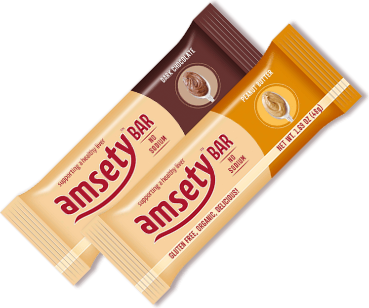
Common Questions
A result below about 5% is generally not considered fatty liver. A result of 5% fat or more defines fatty liver. A small amount of fat in the liver is normal, and a higher percentage means more fat. MRI-PDFF gives one number, so your doctor can compare it with future scans. Your doctor reads the number with your health history, blood tests, and other imaging, so ask what it means for you.
For measuring fat, yes. MRI-PDFF is the most accurate noninvasive method for quantifying liver fat, and it agrees closely with biopsy findings. The CAP score from FibroScan also grades fat well. A score near 275 dB/m suggests fatty liver, but BMI and probe type can affect it. FibroScan is quicker and more widely available, and it also measures liver stiffness, which MRI-PDFF does not. Your doctor chooses the test based on your question and what is available near you.
No. MRI-PDFF measures fat only. It does not measure scarring, inflammation, or how well the liver works. Scarring (fibrosis) is assessed with tests such as MR elastography, FibroScan, or blood-based scores like FIB-4. Some scores, such as the MAST score, combine MRI-PDFF with MR elastography and a blood enzyme to estimate the chance of significant fibrosis or MASH.
Yes, it is considered safe. MRI uses magnets and radio waves and produces no radiation. It carries fewer risks than X-rays and invasive biopsy. Some liver MRI exams use an injected contrast dye, and your care team will tell you if yours does. Tell staff about any implanted devices or metal in your body before the scan. You will also need to stay very still while the scanner records images, so the pictures are clear.
Not in every case. MRI-PDFF measures fat, but a biopsy is still the gold standard for diagnosing MASH, because it shows fat, inflammation, and scarring under a microscope. Biopsy is reserved for an uncertain diagnosis, suspected advanced fibrosis, or clinical trial eligibility. Because a biopsy is invasive, doctors often start with noninvasive tests such as MRI-PDFF. Ask your doctor whether a biopsy is needed in your case.
Medical disclaimer: LiverDirectory provides educational information and a directory of providers. It does not provide medical advice, diagnosis, or treatment. Always consult a licensed physician regarding any medical condition.