AI Summary: HCC imaging surveillance is a routine scan that looks for liver cancer early, before symptoms start, in people at higher risk, most often those with cirrhosis. Ultrasound, usually paired with an AFP blood test, is the standard scan and is repeated about every six months. CT or MRI is used when ultrasound is unclear or a spot is found. A spot is not always cancer. Your doctor sets your schedule and explains results.
Source: www.liverdirectory.com
HCC Imaging Surveillance
Medically Reviewed.
HCC imaging surveillance means regular scans to look for hepatocellular carcinoma (HCC), the most common type of liver cancer, before it causes symptoms. Doctors use it for people whose liver disease raises their risk, most often those with cirrhosis.
Ultrasound is the standard scan, usually paired with an AFP blood test. CT or MRI steps in when ultrasound cannot see the liver well, or when a scan finds a spot that needs a closer look.
This page explains who should be screened, how often, how the scans compare, what results can mean, and how to prepare.
Aimy answers from LiverDirectory’s reviewed health information — helping patients make sense of liver-health terms, prepare for specialist visits, and explore what to look up next.
Surveillance is not the same as diagnosis. It is a routine check for people who have no known liver cancer but a higher chance of developing it. The goal is to find a small tumor early. Early-stage HCC has a much better outlook and more treatment options than advanced disease, according to AASLD guidance on liver nodules.
Ultrasound is the usual first scan. EASL and APASL guidelines call for an ultrasound about every six months in people with cirrhosis. AASLD-based teaching materials say that pairing ultrasound with an AFP blood test is preferred over ultrasound alone. When the ultrasound image is poor, contrast-enhanced CT or MRI is recommended instead.
Your schedule is personal. AASLD guidance weighs the benefits and harms of testing for each person, so your plan may differ from someone else's.
Who needs it
Who Should Have HCC Imaging Surveillance?
Groups Guidelines Include
Adults with Child-Turcotte-Pugh A or B cirrhosis from any cause.
People with Child-Turcotte-Pugh C cirrhosis who are candidates for a liver transplant.
Men age 40 and older and women older than 50 who have chronic hepatitis B without cirrhosis and come from regions where it is common.
Adults with cirrhosis from metabolic fatty liver disease (MAFLD), where APASL puts the yearly HCC rate at about 1% to 1.5%.
People with cirrhosis whose hepatitis C has been cured. EASL advises lifelong regular surveillance for them.
Each scan has a different job. Your care team picks the one that fits your body, your risk, and the question being asked.
HCC Surveillance Ultrasound
Ultrasound is the standard scan for routine surveillance. It uses sound waves, so there is no radiation, and it is painless. A visit usually takes about 30 minutes. Extra body fat or gas in the bowel can make the pictures harder to read.
A multiphase CT scan uses X-rays and contrast dye to photograph the liver in several timed phases: arterial, portal venous, and delayed. Doctors use it when ultrasound is unclear, or to look closely at a spot that ultrasound found. It involves some radiation.
MRI uses magnets and radio waves, not radiation. With contrast dye, it shows how a spot takes up and clears the dye, which helps separate cancer from harmless nodules. It takes longer than ultrasound and needs you to lie still inside the scanner.
Every 6 months: the usual surveillance interval for people with cirrhosis is an ultrasound, often paired with an AFP blood test.
1% to 8% per year: about this share of people with cirrhosis develop HCC each year, depending on the cause, according to a cirrhosis poster cited by Amsety. The cumulative risk over ten years can reach 10% to 30%.
Every 4 months: APASL guidance advises a shorter interval for people who have had HCC before, using ultrasound plus tumor markers.
CT or MRI: AASLD-based teaching materials recommend contrast-enhanced CT or MRI when ultrasound cannot see the liver well.
Two groups are usually not screened: AASLD advises against surveillance in Child-Turcotte-Pugh C cirrhosis when a transplant is not possible, and when other illness limits life expectancy to under one to two years.
How Ultrasound, CT, and MRI Compare
A plain comparison of the three scans. Your doctor decides which to order.
Scan
How it works
Typical role in HCC care
Main limits
Ultrasound
Sound waves; no radiation
Routine surveillance, usually every 6 months
Harder to read with extra body fat or bowel gas
Triple-phase CT
X-rays with contrast dye in several phases
Follow-up when ultrasound is poor or a spot is found
Uses radiation; contrast dye can cause nausea in some people
MRI with contrast
Magnets and radio waves with contrast dye
Follow-up and diagnosis of spots; no radiation
Longer scan; abbreviated MRI lacks enough long-term data to replace ultrasound for routine screening, says EASL
EASL notes that adding AFP to ultrasound raises sensitivity for early HCC but lowers specificity, so more results need a second look.
Results
Understanding Your Surveillance Results
No Concerning Finding
A normal scan is reassuring, but it does not end your care. Surveillance works by repeating. Keep your next appointment on the calendar, and tell your doctor about new symptoms between scans.
A Spot or Nodule Is Found
A spot on ultrasound does not mean cancer. Many nodules in a cirrhotic liver are harmless. Your doctor will usually order a contrast-enhanced CT or MRI to look at the spot more closely.
LI-RADS Categories
Radiologists use a system called LI-RADS to describe spots in people at risk. LR-3 means an intermediate chance of HCC. LR-4 means probable HCC. Neither has the full look of cancer, and LR-4 carries a higher risk than LR-3. AASLD advises close follow-up for these indeterminate nodules, generally with contrast-enhanced imaging every 3 to 6 months.
When Imaging Can Confirm HCC
On a multiphase CT or MRI, HCC typically lights up brightly in the arterial phase, called arterial phase hyperenhancement, and then fades in the portal venous or delayed phase, called washout. In people with cirrhosis, chronic hepatitis B, or past HCC, AASLD and EASL say these features can confirm HCC without a biopsy. EASL says other people need a biopsy, because imaging alone is less reliable for them.
If the Picture Is Still Unclear
If a spot stays suspicious after MRI, EASL says a biopsy should confirm it. AASLD advises against using AFP alone, or liquid biopsy, to diagnose HCC. If cancer is confirmed, a liver cancer team will discuss treatment options with you.
Amsety Bars were designed for people with liver conditions by liver health experts. They offer a convenient way to add energy and nutrients to a liver-friendly diet. Ask your provider what fits your needs.
If you have cirrhosis or another high-risk liver condition, these steps help you get the most from surveillance.
Ask your doctor whether you are due for HCC surveillance and how often.
Ask which scan is planned, and why that one fits you.
Ask whether an AFP blood test will be done with each scan.
Tell the imaging team about allergies, kidney disease, pregnancy, or metal implants before a CT or MRI with contrast dye.
Follow the fasting or diet instructions you are given for each scan.
Keep a list of your scan dates and results, and bring it to visits.
Ask who will call you with results and what happens if a spot is found.
A liver specialist can set a schedule that matches your cause of liver disease and your stage. [Explore liver biopsy](/liver-tests/invasive-testing/liver-biopsy) if your doctor mentions it as a next step.
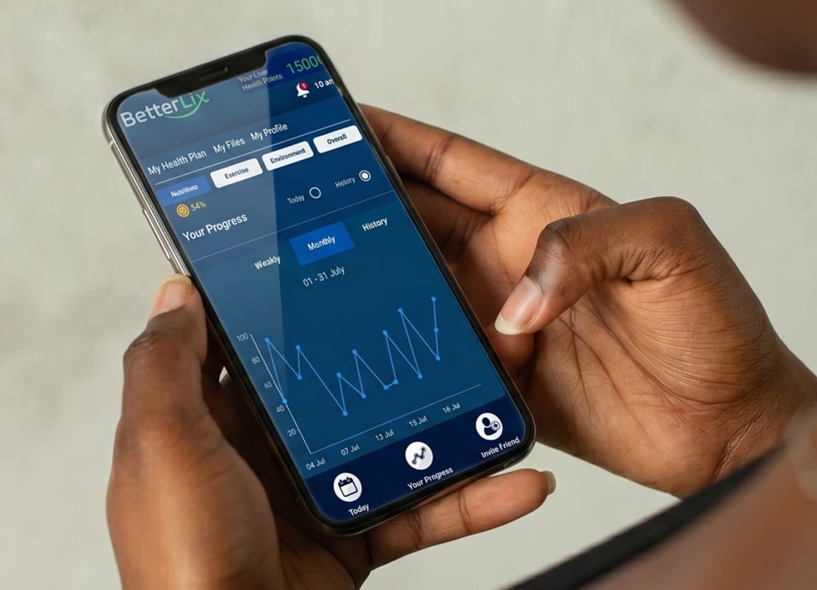
BetterLix is a digital liver health support app designed to help you understand liver conditions, track symptoms, and build healthier daily habits. It provides simple, personalized guidance to help you stay informed about your liver health and monitor progress over time.
Answers to Common HCC Imaging Surveillance Questions
What is HCC imaging surveillance?
HCC imaging surveillance is a routine scan, usually an ultrasound, to look for liver cancer before symptoms start. It is for people at higher risk, most often those with cirrhosis. Guidelines call for a scan about every six months.
How often should I have a liver cancer screening scan?
Every six months for most people with cirrhosis. APASL advises every four months for people who have had HCC before. Your specialist sets your schedule based on your liver disease and stage.
Which scan is best for liver cancer surveillance?
Ultrasound is the standard first scan, and AASLD-based teaching materials prefer pairing it with an AFP blood test. If the ultrasound image is poor, contrast-enhanced CT or MRI is recommended instead.
Does a spot on my ultrasound mean I have liver cancer?
No. Many nodules in a cirrhotic liver are not cancer. Your doctor will usually order a contrast-enhanced CT or MRI, and radiologists rate the spot with LI-RADS. AASLD advises close follow-up for uncertain nodules, generally every 3 to 6 months.
Can ultrasound miss liver cancer?
Sometimes. Ultrasound can be harder to read when there is extra body fat or gas in the bowel. In that case, AASLD-based teaching materials recommend contrast-enhanced CT or MRI. EASL says abbreviated MRI needs more long-term data before it replaces ultrasound for routine screening.
Liver health updates
Stay Updated on Liver Health Insights
Get regular updates on liver diseases, symptoms, prevention tips, and new treatment insights delivered to your inbox.
Medical disclaimer: LiverDirectory provides educational information and a directory of providers. It does not provide medical advice, diagnosis, or treatment. Always consult a licensed physician regarding any medical condition.