AI Summary: Ultrasound-based liver imaging uses sound waves, not radiation, to picture the liver, its blood vessels, and nearby organs. A standard liver ultrasound checks size, shape, and texture and is often the first test when liver disease is suspected. Doppler ultrasound adds blood-flow images, and contrast-enhanced ultrasound (CEUS) gives a closer look at a lesion. A standard exam is painless, takes about 30 minutes, and typically needs no special preparation.
Source: www.liverdirectory.com
Ultrasound-Based Imaging
Medically reviewed content.
Ultrasound-based tests use sound waves, not radiation, to picture the liver, its blood vessels, and nearby organs. This page covers the standard liver ultrasound, Doppler (vascular) ultrasound, and contrast-enhanced ultrasound (CEUS), and links to a detailed page for each.
These tests are noninvasive, widely available, and generally inexpensive compared with CT or MRI, which is why doctors often order one first when liver disease is suspected or when a known condition needs monitoring over time.
Because ultrasound carries no known risk to the patient, it can be repeated as often as a doctor needs, whether that means an initial look at the liver, ongoing surveillance for at-risk patients, or a closer look at a finding from an earlier scan.
Aimy answers from LiverDirectory’s reviewed health information — helping patients make sense of liver-health terms, prepare for specialist visits, and explore what to look up next.
Ultrasound-based imaging uses high-frequency sound waves, not radiation, to create pictures of the liver. A small probe placed on the skin sends sound waves into the body, and a computer turns the reflected waves into images that show the liver's size, shape, and texture. This can reveal changes from a normal liver, including abnormal masses, and often extends to nearby organs such as the gallbladder, bile ducts, and kidneys. The group covers three related tests: standard liver ultrasound, Doppler (vascular) ultrasound, and contrast-enhanced ultrasound (CEUS), each detailed on its own page below.
Key Facts About Ultrasound-Based Liver Imaging
A liver ultrasound images the liver, gallbladder, bile ducts, and nearby organs using sound waves, with no radiation or needles involved.
Doppler, or vascular, liver ultrasound adds blood-flow images to a standard scan and is a primary tool for checking the portal vein and other liver blood vessels.
Contrast-enhanced ultrasound (CEUS) uses an injected microbubble contrast agent, not a dye, to get a clearer view of a lesion already seen on a standard ultrasound.
A standard liver ultrasound is painless and non-invasive, and the exam usually takes about 30 minutes.
Each of these tests builds on the same basic ultrasound technology, but they answer different clinical questions.
Liver Ultrasound (Standard)
A standard liver ultrasound, sometimes called an upper right quadrant or abdominal ultrasound, checks the liver's size, shape, and texture along with the gallbladder, bile ducts, right kidney, and pancreas. Doctors order it after abnormal blood test results or a physical exam finding, or when they suspect liver damage or disease.
A Doppler, or vascular, liver ultrasound combines standard ultrasound images with Doppler readings of blood cell speed and direction. It is a primary, radiation-free tool for evaluating blood flow in the portal vein and other liver vessels, including detecting portal vein thrombosis.
CEUS is ordered when a standard ultrasound spots a lesion that needs a closer look. A provider injects a contrast agent made of tiny gas bubbles, which travels to the liver in the bloodstream and reflects sound waves, making the lesion easier to see and characterize.
Amsety Bars were designed for people with liver conditions by liver health experts. They offer a convenient way to add energy and nutrients to a liver-friendly diet. Ask your provider what fits your needs.
Doctors order these tests for different reasons, from a first look at the liver to monitoring a known vascular problem.
Suspected Liver Damage or Disease
Doctors recommend a standard liver ultrasound when abnormal blood test results or a physical exam suggest liver damage or disease, or when someone has unexplained abdominal pain, enlargement, or distention. It can also be used alongside AFP blood testing as part of liver cancer surveillance, checked every six months in people at higher risk.
Checking Blood Flow Through Liver Vessels
A Doppler (vascular) ultrasound is used when a doctor needs to see how blood moves through the portal vein and other liver vessels. It is the recommended first-line test for diagnosing Budd-Chiari syndrome (hepatic vein obstruction) because it is widely available, low-cost, and a strong screening tool, and it is also used to monitor known portal vein thrombosis over time.
Getting a Closer Look at a Lesion
Contrast-enhanced ultrasound (CEUS) is recommended after a standard ultrasound finds a mass or lesion that needs further characterization. It is also considered when ultrasound visualization is limited and a provider needs more detail without moving straight to a contrast CT or MRI.
Your doctor may ask you to follow a low-fat diet for 24 hours before a liver ultrasound, since the liver's role in digesting fats can affect image quality. Tell your provider about any medications or health conditions beforehand.
During the Exam
A technician applies a water-based gel to your abdomen and moves a probe over the skin, sometimes asking you to shift position for better views. The exam is painless and done on an outpatient basis. For CEUS, a provider injects the contrast agent into a vein before scanning.
Liver health updates
Stay Updated on Liver Health Insights
Get regular updates on liver diseases, symptoms, prevention tips, and new treatment insights delivered to your inbox.
Liver Health Coach
Liver health support
Support Your Liver Health Journey with BetterLix
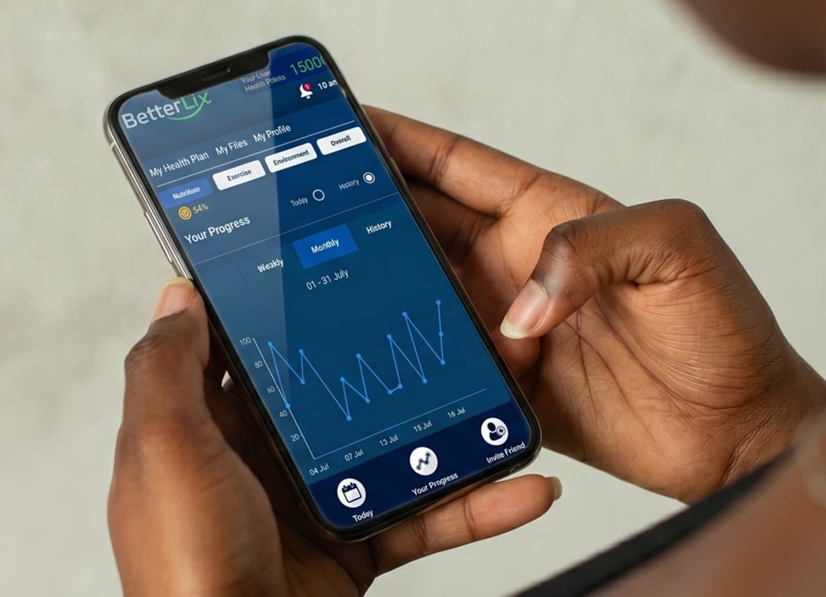
BetterLix is a digital liver health support app designed to help you understand liver conditions, track symptoms, and build healthier daily habits. It provides simple, personalized guidance to help you stay informed about your liver health and monitor progress over time.
Answers to Common Ultrasound-Based Imaging Questions
What is the difference between a liver ultrasound, a Doppler ultrasound, and a CEUS?
A liver ultrasound checks the liver's size, shape, and texture and can spot abnormal masses. Doctors also use special versions: a Doppler (vascular) ultrasound looks at blood flow through liver vessels, and a contrast-enhanced ultrasound (CEUS) gets a closer look at a lesion already found on a standard scan.
Does a liver ultrasound use radiation or contrast dye?
No. A standard liver ultrasound and a Doppler ultrasound use only sound waves and involve no radiation and no contrast agent. A contrast-enhanced ultrasound (CEUS) uses an injected agent made of tiny gas bubbles, not a radioactive dye, and still involves no radiation.
How do I prepare for a liver ultrasound?
Your doctor may recommend a low-fat diet for about 24 hours beforehand, since digesting fat can affect image quality. Otherwise, no special preparation is typically needed for a standard liver ultrasound.
Is a liver ultrasound painful or risky?
A standard liver ultrasound is painless and non-invasive. You may feel light pressure from the probe and a water-based gel on your skin, and a CEUS adds a brief injection, but there are no reported risks associated with the procedure itself.
When do I get my liver ultrasound results?
Results can be instant: the physician performing or reviewing the scan may point out findings right away. If the images need more review, your doctor will share the results with you afterward, and may recommend further testing, such as an MRI, CT scan, or biopsy, if the ultrasound is unclear.
Medical disclaimer: LiverDirectory provides educational information and a directory of providers. It does not provide medical advice, diagnosis, or treatment. Always consult a licensed physician regarding any medical condition.